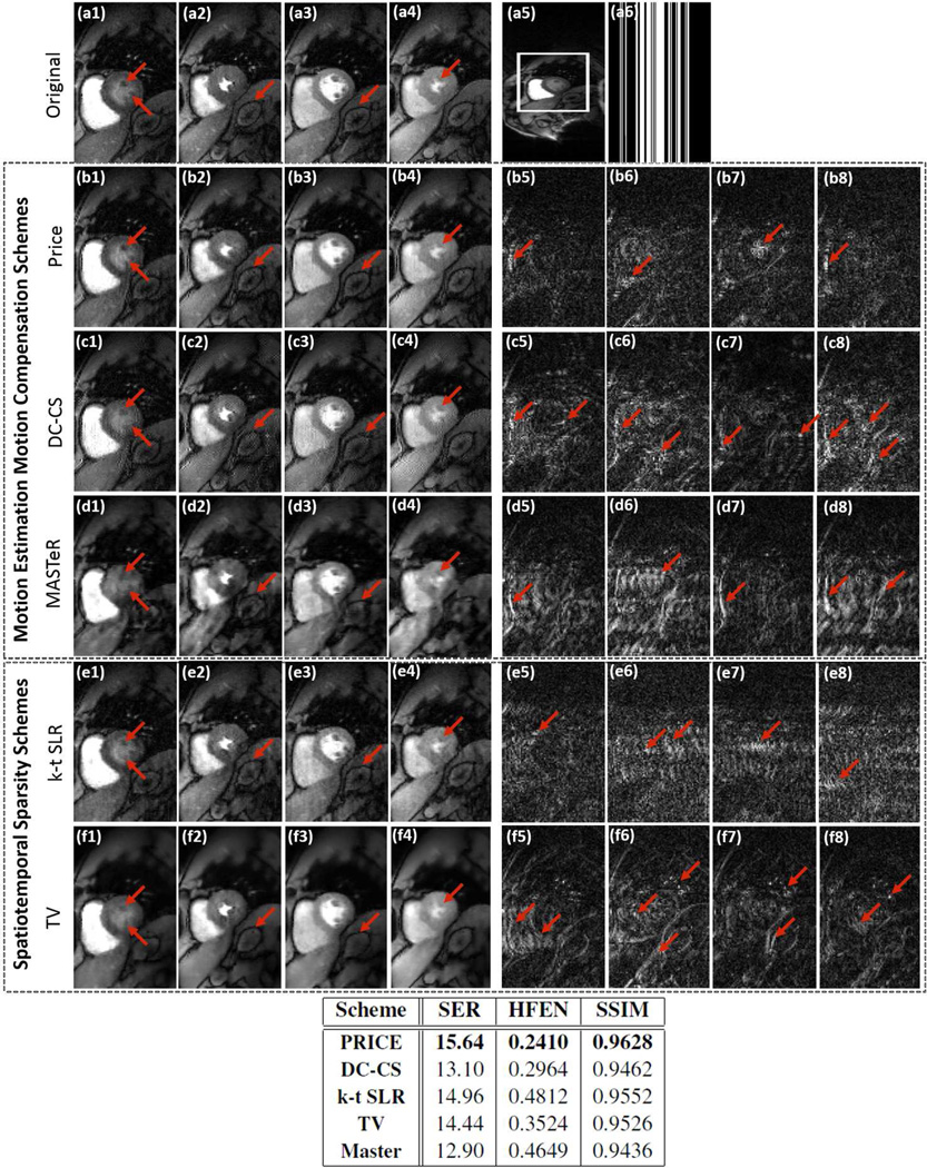
Fig 4.
Evaluation of the ME-MC algorithms by retrospectively downsampling ungated & free-breathing myocardial perfusion MRI data. The images (a1)–(a4) correspond to frames in the time series with different cardiac/respiratory phases and different contrast due to bolus passage. These images are cropped from a 288×108×80 dataset acquired with 6 coils; one of these images are shown in (a5). We undersampled the Cartesian sampled data along the phase encoding direction to obtain a three-fold acceleration. One of the sampling masks are shown in (a6). The reconstructions and corresponding residuals using PRICE (b1–b4) & (b5–b8), DC-CS (c1–c4)& (c5–c8), MASTeR (d1–d4)& (d5–d8), k-t SLR (e1–e4)& (e5–e8) and TV (f1–f4)& (f5–f8) are shown. The error images are scaled by a factor of three for better visualization. The extensive inter-frame motion and contrast variations due to bolus passage makes this dataset very challenging. We observe that the PRICE scheme provides reconstructions with lower spatial and temporal blurring, compared to the other algorithms. The table above shows a quantitative comparison of the entire methods using SER, HFEN and SSIM metrics computed on the region of interest shown in (a5).

