Figure 2.


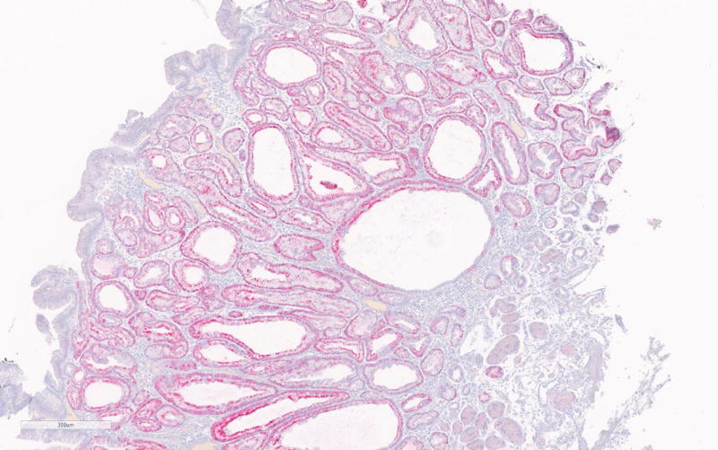
a. MUC6 immunohistochemistry of PGA showing strong positivity of the glandular compartment.
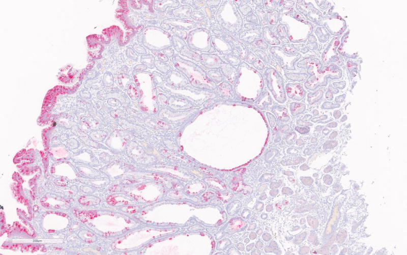
b. MUC5A immunohistochemistry showing positivity of the foveolar epithelium.
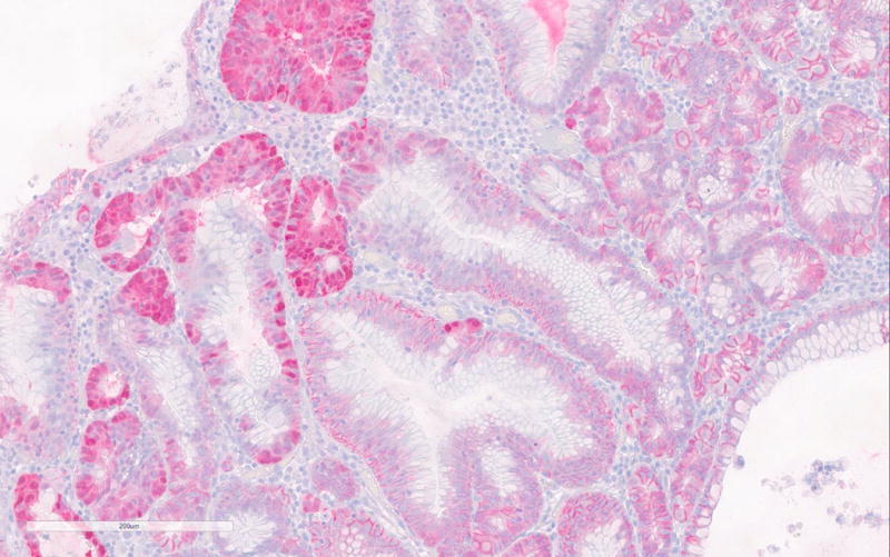
c. Nuclear β-catenin expression in areas with high-grade dysplasia in PGA3.
