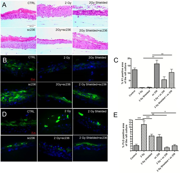
Figure 3.
Rescue of the normal morphology of the 3D cultures after irradiation in the presence of 5 µmol/l specific cyclooxygenase-2 inhibitor sc-236 7 days postirradiation. (A) H&E staining; immunofluorescence staining of (B) cytokeratin 1 and filaggrin (D) as differentiation markers. Quantification of the differentiation maker expression by Image J as described in Section “Materials and Methods.” (C) and (E) Blue—DAPI; green—K1. Error bars—SEM; **p < 0.01; ***p < 0.001, one-way ANOVA analysis, Tukey posttest.

