Figure 1.
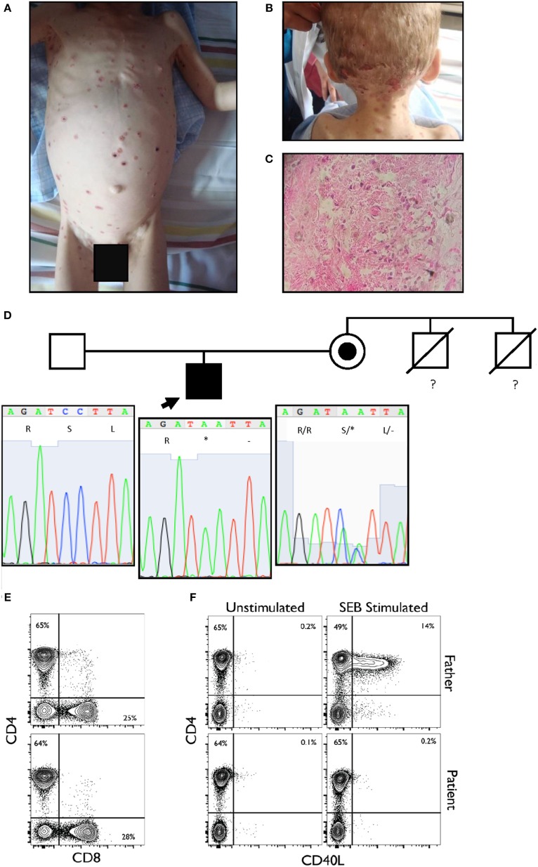
(A,B) Disseminated vesicular or dry erythematous and ulcerous lesions over the body and scalp of the proband. (C) Histological analysis of the dermis showed intense edema, necrosis of the dermal fibers, and pseudo-granulomatous tissue. A few giant cells and a polymorphonuclear infiltrate are observed. Macrophages are present, loaded with oval-shaped parasites, between 2 and 4 µm in size, positive for periodic acid–Schiff staining (intense red-violet), consistent with Histoplasma capsulatum. (D) Pedigree of the family sequenced in this study. The arrow indicates the proband. The maternal uncles died of unspecified infections before they reached two years of age. The lower panel shows the Sanger sequencing results and familial segregation for the mutation in CD40LG in the affected proband and both unaffected parents. The mutation is inherited from the mother. (E) Normal CD4 and CD8 percentages and ratio within the CD3+ population. (F) Complete absence of CD40L upregulation in patient CD3+ population when stimulated for 6 h with SEB as compared to healthy paternal sample.

