Figure 6.
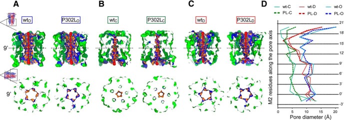
The pore mutation P302L destabilized the channel gate in the open state. A-C, Visualization of pore sections in the open (A), closed (B), and desensitized (C) states were shown parallel to the membrane (top panels) and from the extracellular side (bottom panels) of wt and P302L structures. In the top panels, the section of each structure was obtained by cutting the protein structure along the pore-axis. Red spheres represented pore centers at given pore heights and their diameters correspond to 1/10 of the pore diameter calculated at that point. Bottom panels represent transverse sections of the pore-axis at the 9’ position (dashed lines in top panels), where the channel gate was located. Pore-lining side chains and residues at 3-Å steps within the cavity were colored in orange and blue, respectively. The remaining part of the transmembrane domain was shown in green. D, Pore diameter profiles at 3-Å steps corresponding to the pore-lining residues in the open (A), closed (B), and desensitized (C) states of wt and P302L structures. Horizontal black dotted lines show positions of pore-lining residues in M2 (see Figure 1A). PL, P302L. Letters in subscript refer to open (O), closed (C), or desensitized (D) states.

