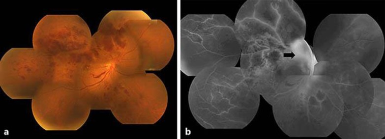
Fig. 2.
Funduscopy (a) and fluorescein fundus (b) images obtained at 50 days after the initial examination. Neovascularization that caused vitreous hemorrhage can be seen from the optic disc to the upper retinal vascular arcade. Fluorescein angiography revealed dye leakage from the optic disc neovascularization and fibrovascular membrane (black arrow), and enlargement of the nonperfusion area in the affected retina. The corrected visual acuity of the right eye had decreased to 0.02.

