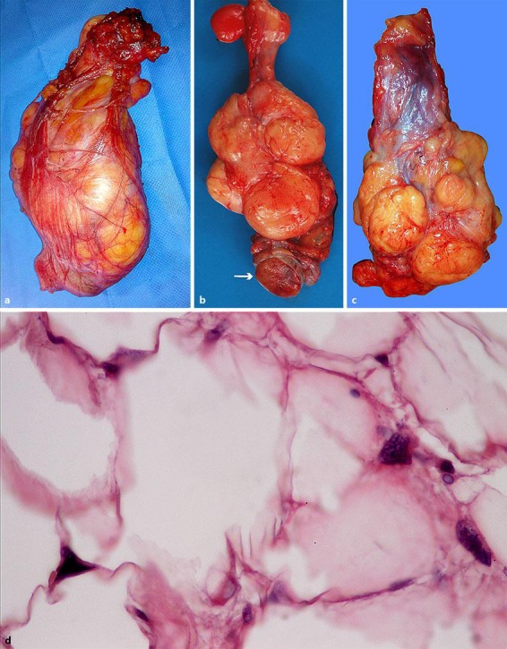
Fig. 2.

The spermatic cord was dissected and removed. a It showed a hard lipomatous mass that was 14 × 8 cm in size. The mass involved the entire circumference of the cord and could not be separated from the cord. b The mass had a “bunch of grapes” appearance and consisted of several masses of various sizes that surrounded the spermatic cord. c Macroscopic examination of the testis showed that it appeared to be normal. d Histopathological examination revealed a well-differentiated liposarcoma of the cord with positive margins; some lipoblasts with indented hyperchromatic nuclei were observed (haematoxylin-eosin staining; magnification ×400).
