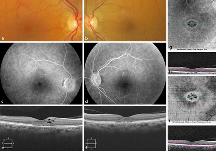
Fig. 1.
Bilateral cystoid macular edema in a 52-year-old woman treated with tamoxifen and docetaxel. Fundus color photograph of the right eye (a) and the left eye (b): stellate appearance of the macula. Late phase of the fluorescein angiogram in the right eye (c) and the left eye (d) shows no dye pooling in cystoid spaces. SD-OCT horizontal B-scans of the right (e) and left eye (f) showing intraretinal cysts and fragmentation of the ellipsoid zone. Right eye En-face OCT (g) and its segmentation (h) in the right inner retina showing the petaloid aspect of cysts in the macula. Right eye En-face OCT (i) and location of its segmentation (j) in the right outer retina showing the petaloid aspect of the outer retina.

