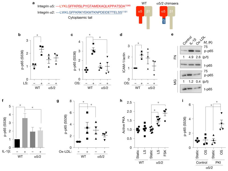
Figure 1.
Integrin α subunit cytoplasmic tails determine ECM-specific inflammatory signalling. (a) Alignment of integrin α subunit tail sequences and schematic representation of integrin α5/2 chimaera. (b,c) NF-κB activation by flow. WT α5 or α5/2 cells on FN (20 μg ml−1) were subjected to 30 min laminar shear (b) or 18 h oscillatory shear (c). NF-κB activation was then assayed by western blotting for Ser536 p65 phosphorylation (b, n = 3; c, n = 4). Y axis values throughout the figure represent the fold change (relative to control). LS, laminar shear; OS, oscillatory shear. (d) Induction of ICAM-1 after 18 h of oscillatory flow in cells expressing WT α5 versus the α5/2 chimaera on FN (n = 3). (e–g) NF-κB activation by soluble atherogenic factors. WT BAECs on FN or diluted Matrigel (MG) were stimulated with IL-1β or oxidized LDL for 30 min. NF-κB was assayed by western blotting for pSer536 p65 (e). t-p65, total p65. Quantification in f (n = 7) and g (n = 4) shows activation relative to control cells on FN without flow. (h) Effect of the chimaera on PKA activation. WT α5 or α5/2 chimaera cells on FN were sheared for 15 min and active PKA was pulled down from cell lysates with GST-PKI followed by immunoblotting. Forskolin (FSK) was used as a positive control for PKA activation (n= 4–8). (i) PKA inhibition rescues NF-κB activation in α5/2 chimaera cells. Chimaera cells on FN were treated with the PKI 14–22 amide inhibitor or dimethylsulfoxide (DMSO) alone, and then subjected to oscillatory shear for 18 h. Activation of NF-κB was assayed by western blotting (n= 4). Data are represented as means ± s.e.m. *P < 0.05 by one-way ANOVA (b,c,h,i) or two-tailed t-test (d,f,g). In all panels n values represent independent experiments. Source data are provided in Supplementary Table 1. Unprocessed scans of blots are shown in Supplementary Fig. 6.

