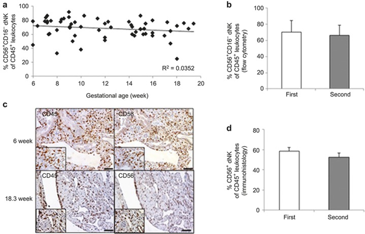
Figure 1.
Quantification of the dNK cell population throughout the first to second trimester of pregnancy. (a) Decidual leukocytes were isolated from the 6- to 20-week pregnancy period. The percentage of viable CD45+CD56+CD16− dNK cells at different gestational ages was illustrated in a scatterplot. Regression trend lines and R2 value are included. n = 65. (b) Histograms summarize the average proportion of first and second trimester dNK cells based on flow cytometric results. n = 34 (first trimester; 9 ± 1.9 week) or n = 31(second trimester; 16 ± 1.5 week). (c) Representative photographs of CD45 and CD56 immunohistochemical staining of serial sections of first and second trimester decidual tissues. High power images were inserted. Scale bar = 100 μm. (d) Summary data of the CD56+ dNK cell proportion amongst the CD45+ leukocytes from immunohistological analysis. n = 19 (first trimester) and 18 (second trimester).

