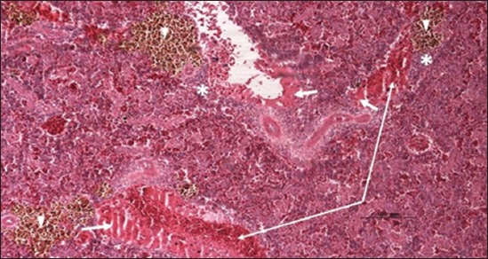
Figure-18.

Spleen parenchyma section (H and E stain) of hybrid tilapia (Oreochromis spp.) naturally infected by Streptococcus agalactiae showing large thrombus in the splenic blood vessel (thick arrow), multifocal hemosiderin deposition (head arrow), congestion of blood vessels (thin arrow), and multifocal infiltration of macrophages (asterisk).
