Figure-6.

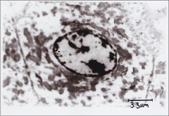
Transmission electron microscopy of hepatocyte showing regenerated mitochondria, rough endoplasmic reticulum, normal nucleus, nucleolus and cytoplasm showing vesicular structures (Group 3).

Transmission electron microscopy of hepatocyte showing regenerated mitochondria, rough endoplasmic reticulum, normal nucleus, nucleolus and cytoplasm showing vesicular structures (Group 3).