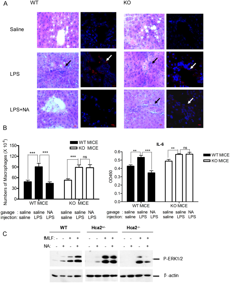
Figure 8. Niacin inhibits LPS-induced acute endotoxemia in mice.
Hca2 KO and WT mice were sacrificed 4 h following endotoxemia induction. (A) HE staining (original magnification, 400×, bar: 10 μM) and immunofluorescent staining by anti-F4/80 antibody (original magnification, 630×, bar: 10 μM) of liver sections from saline-, LPS- or LPS + niacin-treated WT and Hca2−/− mice. (B) The number of macrophages isolated from the ascites of WT and Hca2−/− mice (n = 10) and IL-6 in the ascites of WT and Hca2−/− mice treated with saline, LPS, or LPS + niacin (n = 8–10). Error bars represent standard deviation of the means (*p < 0.05; **p < 0.01, ***p < 0.001, compared with the indicated group). (C) ERK1/2 activation in niacin-treated macrophages from WT, Hca2+/− and Hca2−/− mice.

