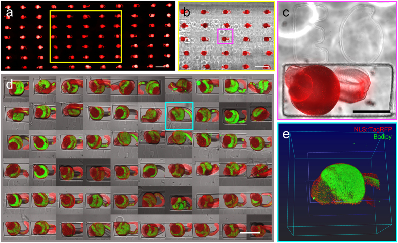
Figure 5. Transgenic zebrafish larva serial imaging in different conditions.
(a–c) Widefield fluorescence and brightfield stitched images of large field of views. a. Stitching of a very large field of view acquired with a 4X objective and reconstructed by the acquisition software. (b) Central zone outlined in yellow in (a) is shown as an overlay of brightfield and fluorescence, this stitched view is acquired with a 10X objective. (c) Crop of the central embryo (magenta square in b) illustrates the resolution of imaging making discernible the single nuclei of each cell of the embryo. (d,e) Confocal imaging of the transgenic embryos counterstained with the lipid marker Bodipy. (d) Tile of the 60 fields of views acquired with confocal microscopy. Overlay of fluorescence projection and brightfield images. (e) 3D rendering of the two fluorescent channels and depiction of the bottom and top of the well (blue lines). All bars are 500 μm.

