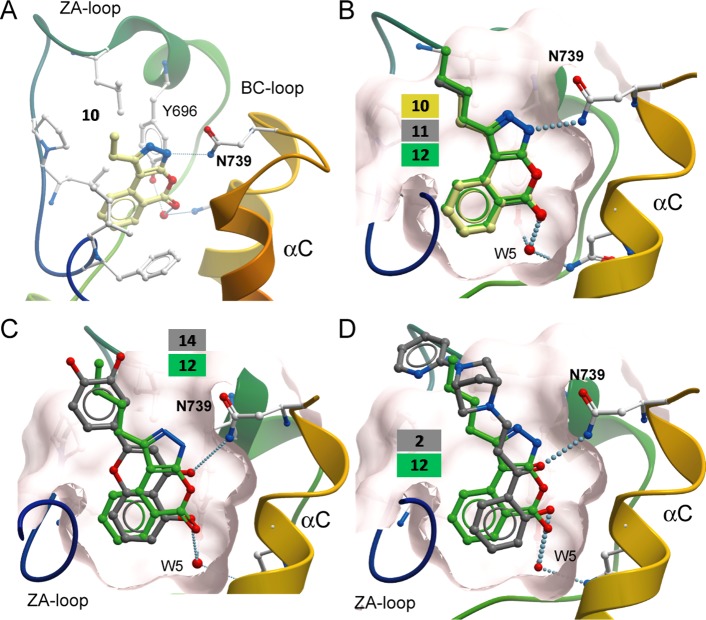
Figure 7.
Binding mode comparison. (A) Binding mode of compound 10 in PB1(5). (B) Superimposition of the binding modes of inhibitors 10–12 showing a high degree of similarity. The remaining conserved water molecule (W5) is highlighted. Hydrogen bonds are indicated by dotted lines. The surface of the acetyl-lysine binding pocket is shown as a transparent sphere. (C) Comparison of binding modes of 12 and 14. (D) Comparison of binding modes of 12 and the PB1/SMARCA inhibitor 2.

