
Fig. 7.
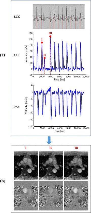
Real-time PC-CMR for the 23-year-old arrhythmic patient. a: The ECG recordings and the velocity waveforms of AAo and DAo. b: The magnitude images and velocity maps for the three representative time frames within an arrhythmic period. As can be seen, the proposed method nicely captures a dramatic change of flow velocities occurring during an arrhythmia period. Note that this type of flow dynamics cannot be obtained from the conventional cine method