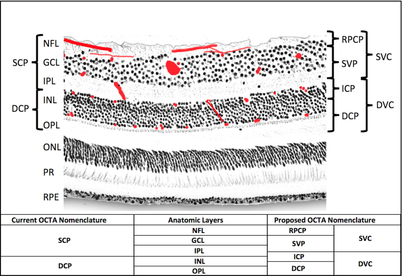
Figure 1. Anatomic localization of vascular plexuses in the human retina in the macula, and current and proposed optical coherence tomography angiography segmentation boundaries.
An illustration of the retinal vascular plexuses in red (labeled on right) hand drawn on top of a histological section of the human retina showing anatomic layers (labeled on left) from spectral domain optical coherence tomography. The four vascular plexuses can be grouped into superficial and deep vascular complexes (SVC and DVC, as shown on right) for routine segmentation, but ought to reflect the anatomic location of the ICP at the IPL/INL interface, which the current OCTA segmentations use as a border between superficial and deep plexuses (labeled on left as SCP and DCP). Current and proposed vascular nomenclature and OCTA segmentations are shown at the bottom. (NFL = nerve fiber layer, GCL = ganglion cell layer, IPL = inner plexiform layer, INL = inner nuclear layer, OPL = outer plexiform layer plus Henle’s fiber layer, ONL = outer nuclear layer, PR = photoreceptor layers, RPE = retinal pigment epithelium, OCTA = optical coherence tomography angiography, RPCP = radial peripapillary capillary plexus, SVP = superficial vascular plexus, ICP = intermediate capillary plexus, DCP = deep capillary plexus).

