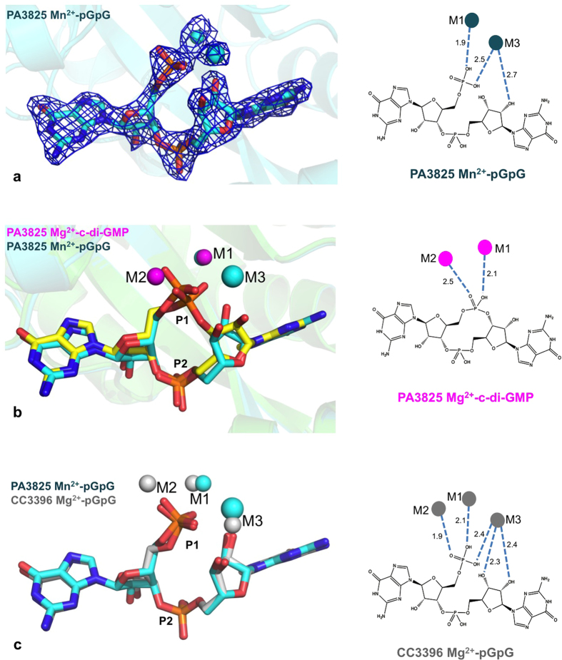
Figure 5. Identification of the M3 position in the pGpG complex.
(a) Electron density of pGpG and metal ions bound to PA3825EAL, contoured at 1.3 σ. (b) Structural overlay of the substrate and product complexes of PA3825EAL. The substrate c-di-GMP is shown as yellow sticks; Mg2+ ions (magenta spheres) occupy the M1 and M2 binding sites. The product pGpG is shown as blue sticks; Mn2+ ions (blue spheres) occupy the M1 and M3 binding sites. (c) Structural comparison of the product complexes of PA3825EAL and CC3396EAL (PDB code 3U2E). In PA3825EAL manganese and sodium ions are present in the M1 and M3 positions, respectively, while in CC3396EAL magnesium ions are present in the M1, M2 and M3 positions. Schematic representations on the right show the interactions between the coordinated metal ions and the bound substrate or product, with distances labelled in Å.

