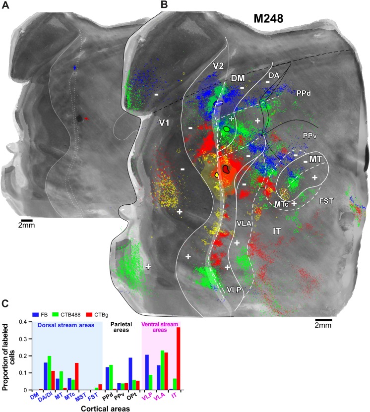
Fig. 6.
A column of four different tracer injections involving upper field DM and cortex medial and lateral to it reveals two distinct areas bordering V2d. Case M248. (A) CO image of unfolded and flattened visual cortex. The pale CO spot (blue arrow) is the location of the FB injection site, whereas the dark spot is the silver reacted CTBg injection site (red arrow). (B) The same CO image as in (A) is shown enlarged with overlaid injection sites and plotted cell label resulting from each tracer injection (FB, blue; CTB488, green; CTBg, red; DY, yellow). The locations of anterogradely labeled fibers are outlined. White arrow on the FB injection site indicates the direction of travel of the injection site from superficial to deeper layers. (C) Proportion of labeled cells in each extrastriate area resulting from the FB, CTB488, and CTBg injections. For abbreviations, see legend of Fig. 1. Other conventions are as in Fig. 3.

