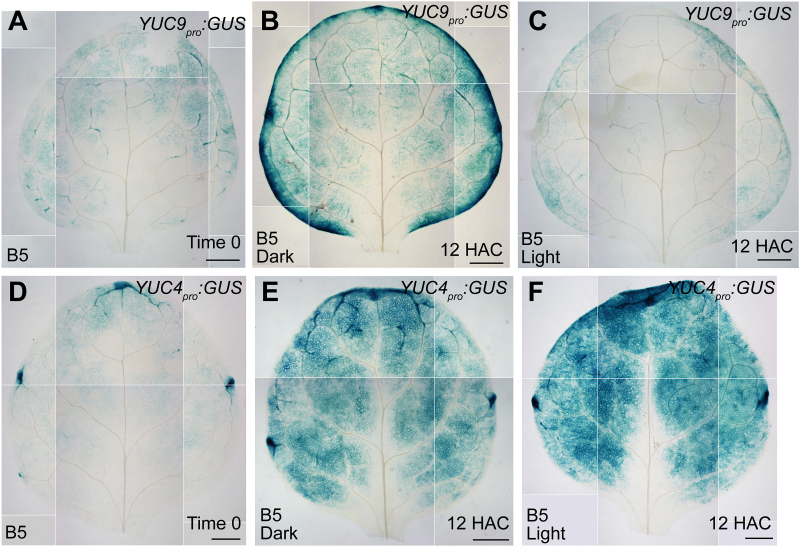
Fig. 8.
Expression patterns of YUC9 and YUC4 in dark and light conditions. (A) GUS staining of the time-0 leaf explant from YUC9 pro :GUS. (B, C) GUS staining of 12-HAC leaf explants from YUC9 pro :GUS cultured on B5 medium in dark (B) or light (C) conditions. (D) GUS staining of the time-0 leaf explant from YUC4 pro :GUS. (E, F) GUS staining of 12-HAC leaf explants from YUC4 pro :GUS cultured on B5 medium in dark (E) or light (F) conditions. The data in A–F were pasted together from small pictures of the same leaf explant because the microscope was unable to capture the entire leaf explant in a single visual field. Scale bars, 500 μm in A–F.

