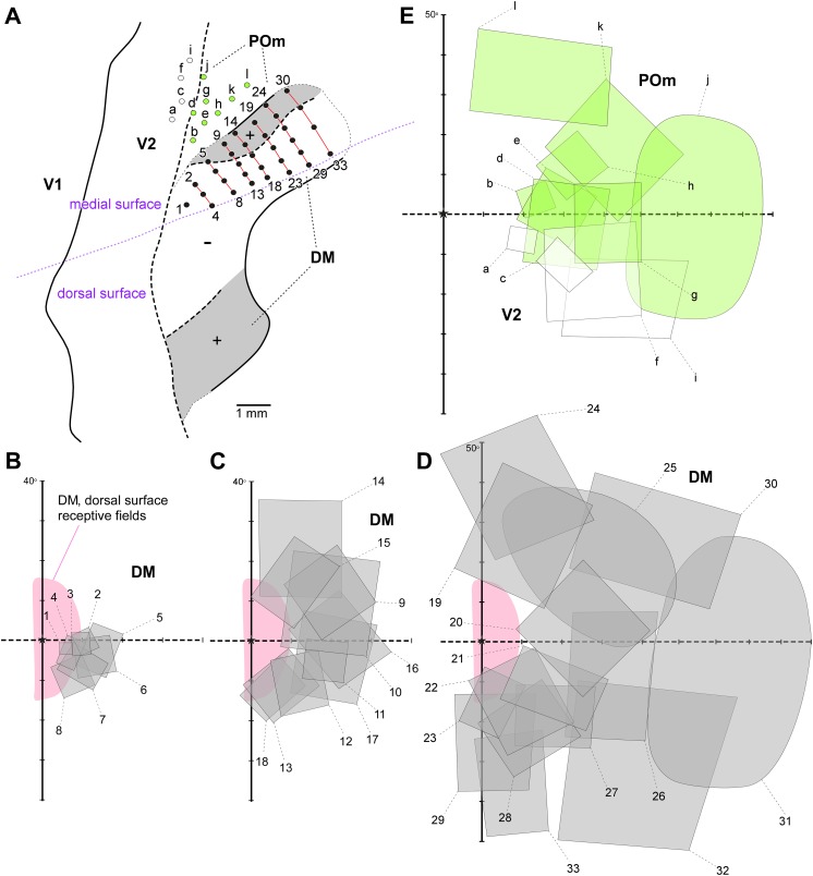
Fig. 6.
The part of area DM located on the midline surface contains the representation of the peripheral visual field. Receptive fields obtained from sites on the medial surface of the occipital lobe, in long tangential electrode penetrations (from Rosa et al., 2005). (A) Diagram of an unfolded map of the cortex, showing the locations of recording sites relative to the borders of V2, DM and another third tier area (POm) that is located adjacent to the representation of the lower far peripheral visual field in V2. The dotted purple line indicates the medial convexity of the occipital lobe. (B–D) Receptive fields in area DM. In each diagram, the region of visual field that is represented on the dorsal surface of the brain of the same animal is shaded in pink. Recording sites and receptive fields in DM are numbered 1–33, and those in V2 and POm are designated by letters. Recording sites and receptive fields in POm are indicated in green, those in V2 in white.

