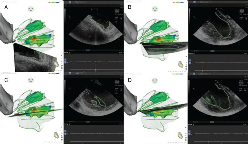
Figure 1.
(A) ICE 3D catheter orientated towards the LV apex. (B) Clockwise rotation of the ICE 3D catheter showing an LV long axis. (C) Posteromedial papillary muscle body and chordae. (D) Posteromedial papillary muscle base. Contours are drawn in the ICE 2D clip and then transpolated to the 3D geometry.

