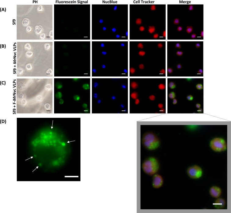
Figure 1. Triple fluorescence labelling and detection of MrNvc VLPs in Sf9 cells.
(A) Sf9 cells in the absence of MrNvc VLPs, (B) Sf9 cells incubated with non-labelled MrNvc VLPs and (C) Sf9 cells incubated with F-MrNvc VLPs. The cell nucleus and cytoplasm were labelled with NucBlue Live Ready Probe Reagent (blue) and Cell Tracker Orange (red), respectively. PH indicates images captured under white light. Merge images represent the superimposed green, blue and red signals. (D) Small granular appearance in a Sf9 cell incubated with F-MrNvc VLPs is indicated by the arrows. Bars, 10 µm.

