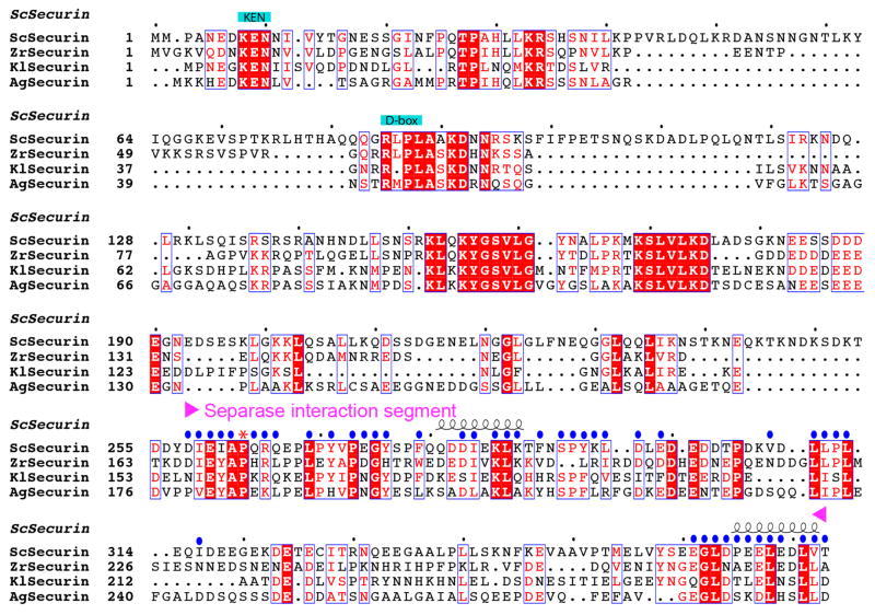
Extended Data Fig. 4.
Sequence alignment of securin. The separase interaction segment (SIS) is indicated. Residues in contact with separase are indicated with the blue dots. Residue 263 is equivalent to the P1 residue of separase substrates, and is indicated with the red asterisk. Residues 317–360 of S. cerevisiae securin are disordered in the current structures and are poorly conserved in sequence.

