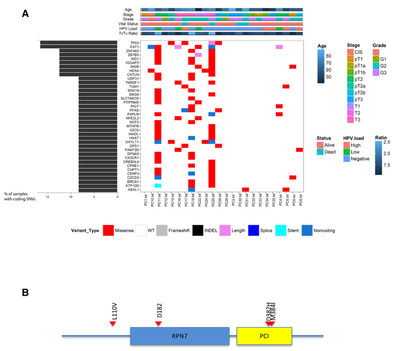
Figure 1. Somatic variants in penile cancer.
A, recurrently mutated genes in penile cancer. The central heatmap shows the mutation status of the recurrently mutated genes for each tumor. Somatic mutations are colored according to functional class (lower). Left, mutation count for each individual gene. Top, patient phenotype data for age, stage grade, Ti/Tv ratio and HPV viral status. B, schematic representation of the CSN1 protein. The conserved RPN7 and PCI domains are shown in blue and yellow, respectively. CSN1 alterations, position, and aa substitution are highlighted. Red, missense mutations.

