
Figure 1.
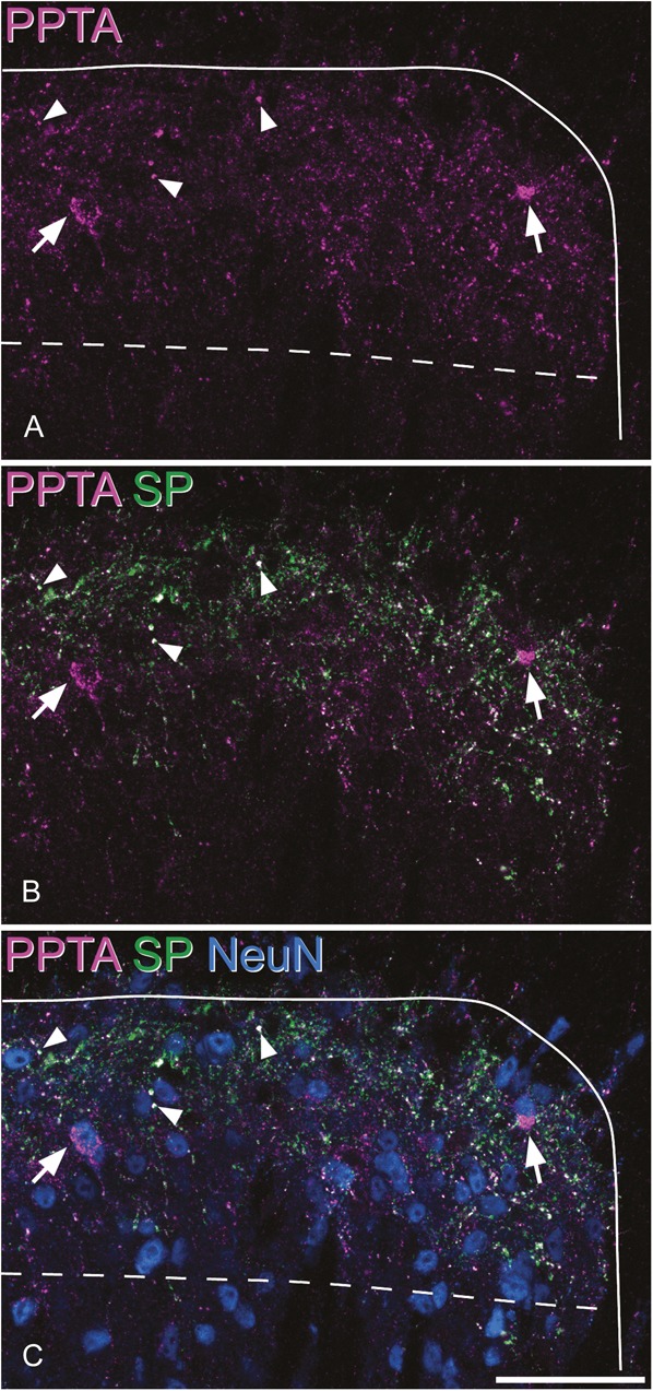
Immunostaining for preprotachykinin A (PPTA), substance P (SP), and NeuN in the mouse dorsal horn. (A) PPTA immunoreactivity (magenta) appears as fine punctate structures, many of which are axons (3 indicated with arrowheads), as well as large clumps (arrows) that represent staining in the perikaryal cytoplasm of certain neurons. (B) SP (green) is colocalised with PPTA in the axons, but not in the perikaryal cytoplasm. (C) Staining for the neuron-specific protein NeuN confirms that the clumps of PPTA shown with the arrows are in the cell bodies of 2 of the neurons. The solid line indicates the outline of the gray matter, and the dashed line shows the border between laminae II and III. The images are projected from 2 optical sections at 1 μm z-spacing. Scale bar = 50 μm.