
Figure 9.
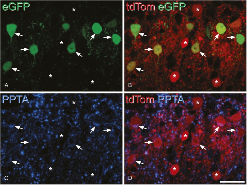
Preprotachykinin A (PPTA) immunoreactivity in enhanced green fluorescent protein (eGFP)–expressing neurons after intraspinal injection of AAV.flex.eGFP into a Tac1Cre;Ai9 mouse. The section has been scanned to reveal eGFP (green), tdTomato (tdTom) (red), and PPTA immunoreactivity (blue). (A) This field shows several eGFP+ neurons, and some are indicated with arrows. (B) All the eGFP+ neurons also express tdTom, and there are additional tdTom+ cells that lack eGFP (some marked with asterisks). (C) Shows PPTA immunoreactivity in the same field, and in (D), this has been merged with tdTom. PPTA can be seen in some of the cells marked with arrows (ie, those that are also eGFP+). It is present in the form of clumps within the perikaryal cytoplasm, where it overlaps the tdTom. Note that the tdTom+/eGFP− neurons are not PPTA immunoreactive. The section is a projection of 3 optical sections at 0.5 μm z-spacing. Scale bar = 20 μm.