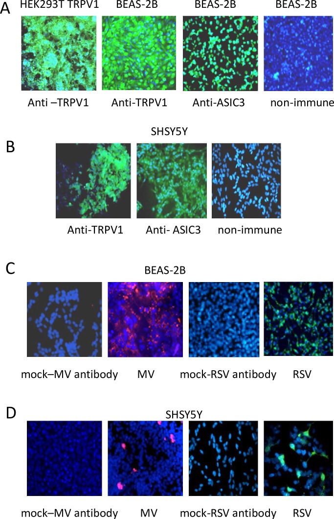
Fig 1. BEAS-2B and SHY5Y5 cells express TRPV1 and ASIC3 and support virus replication.
(A) BEAS-2B and (B) SHSY5Y cells were stained with anti-TRPV1, anti-ASIC3 antibodies or non-immune rabbit serum and DAPI. HEK293T TRPV1 transfected cells were used as positive control for TRPV1 expression. Receptors (green), nuclei (blue). (C) BEAS-2B cells were mock infected or infected with RSV or MV at an MOI of 1 for 48 (D) SHSY5Y cells were mock infected or infected with RSV or MV at an MOI of 1 for 72 hours and stained with anti-viral antibodies and DAPI. RSV (green), MV (red), nuclei (blue). Magnification X 200.

