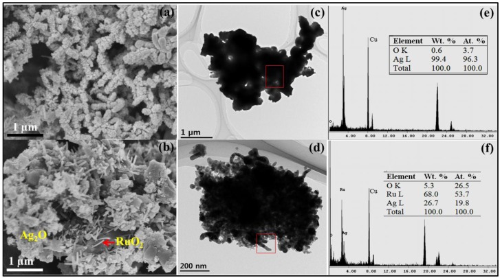
Figure 2.
Field emission scanning electron microscope (FESEM) surface morphological images of (a) Ag2O nanomaterials (NMs); and (b) Ag2O/RuO2; high resolution transmission electron microscope (TEM) images of (c) Ag2O NMs; and (d) Ag2O/RuO2; and energy dispersive X-ray spectroscopy (EDX) spectra with the elemental composition for (e) Ag2O NMs; and (f) Ag2O/RuO2.

