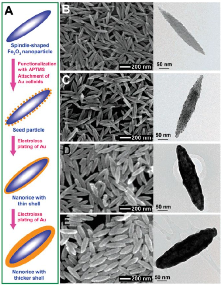
Figure 6.
(A) Schematics of the fabrication of hematite-Au core-shell nanorice particles. SEM (left) and transmission electron microscopy (TEM) (right) images of (B) hematite core (longitudinal diameter of 340 ± 20 nm, and transverse diameter of 54 ± 4 nm; (C) seed particles; (D) nanorice particles with thin shells (13.1 ± 1.1 nm); and (E) nanorice particles with thick shells (27.5 ± 1.7 nm). Reproduced with permission from [73]. Copyright American Chemical Society, 2006.

