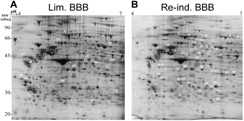
Figure 2.
Comparison of the proteins extracted with Triton X-100 from bovine brain capillary endothelial cells showing limited (Lim. BBB) (A) or re-induced (Re-ind. BBB) BBB functionalities (B). Digital image obtained after 2D-PAGE of the proteins separated according to their pI and MW. The gel was silver nitrate stained. The numbering corresponds to the enriched protein in each condition. Each spot was identified by peptide mass fingerprinting (PMF) and/or peptide fragmentation fingerprinting (PFF) on a Proteineer TM workstation (adapted with permission from [45]).

