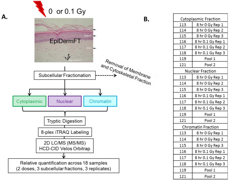
Figure 2.
Experimental design for 8-plex iTRAQ experiment. (A) Reconstituted skin tissues were separated into subcellular fractions 8 h post-exposure to 0 or 0.1 Gy ionizing radiation. Each fraction was digested with trypsin, labeled with 8-plex iTRAQ reagents, recombined and analyzed using 2D LC/MS; (B) Each 8-plex analysis contained three biological replicates (Rep) of 0 and 0.1 Gy-treated subcellular fraction. Pooled fractions were included to facilitate comparisons across samples.

