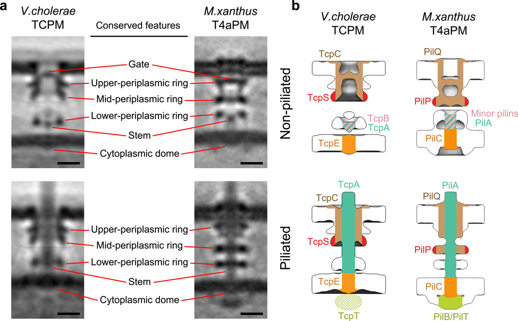
Figure 2. Comparison between V. cholerae TCPM and M. xanthus T4aPM structures, and the inferred TCPM component locations based on the T4aPM component map.
(a) Left column: slices through the composite sub-tomogram averages of V. cholerae wild-type non-piliated and piliated TCPM structures. Right column: slices through sub-tomogram averages of M. xanthus ΔpilB non-piliated and wild-type piliated T4aPM structures8. Scale bars, 10 nm. (b) Inferred TCPM component locations of TcpC, TcpS, TcpB, TcpA and TcpE (left column) based on their identified analogy to PilQ, PilP, minor pilins, PilA and PilC, respectively, in the reported T4aPM component map8 (right column).

