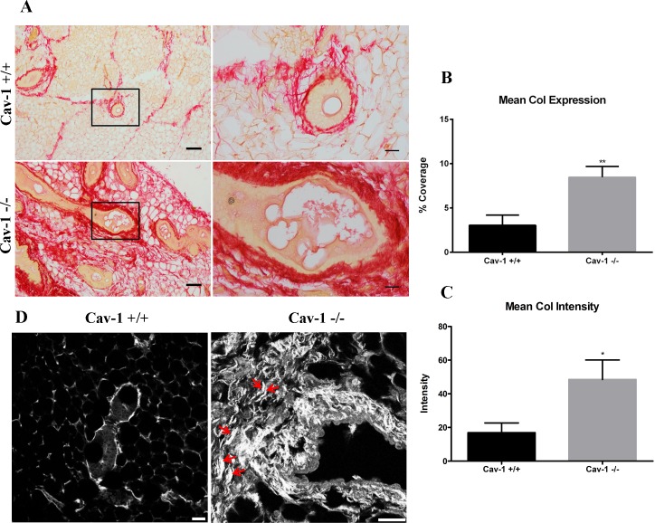
Fig 3. Collagen I expression and organization are upregulated in cav-1 -/- mammary glands.
(A) PSR staining for collagen I in cav-1 +/+ and cav-1 -/- glands at low and high magnification. Imaging demonstrates extensive expression of collagen I in the adipose stroma and in the peri-ductal region of cav-1 -/- glands. The collagen I fibers appear dense and tightly packed in cav-1 -/- glands. In contrast, overall collagen I expression is considerably lower in cav-1 +/+ glands with fibers being predominantly located to the peri-ductal region. Here, fibers are less packed and distinct fibrils are apparent. (B) The percent expression of PSR stained collagen I was significantly higher in cav-1 -/- glands compared to cav-1 +/+ glands. Scale bars are 50 μM and 20 μM in low and high magnification images, respectively. (C) The intensity of PSR stained collagen I was significantly higher in cav-1 -/- glands compared to cav-1 +/+ glands. (D) Representative SHG images show an abundance of dense, thick collagen fibers in cav-1 -/- glands. Collagen fibers and fiber bundles also appear more organized and aligned (red arrows). Cav-1 +/+ glands express comparably less collagen which presents as thin, unorganized fibers. Scale bars are 25 μM. Col: collagen. *p≤0.05, **p≤0.01. Results are reported as the mean ± the SEM.

