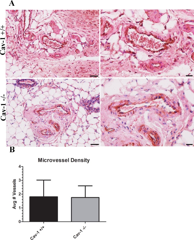
Fig 5. Microvessel density is similar in cav-1 -/- and cav-1 +/+ glands.
(A) Immunohistochemistry of CD31 expressing endothelial cells in cav-1 +/+ and cav-1 -/- glands at low and high magnification. Results demonstrated that vessels are apparent in both cav-1 +/+ and cav-1 -/- glands. (B) Quantification of microvessels show no differences in the abundance of vessels in cav-1 +/+ and cav-1 -/- glands. Scale bars are 50μM and 20μM for low and high magnification images, respectively. Results are reported as the mean ± the SD.

