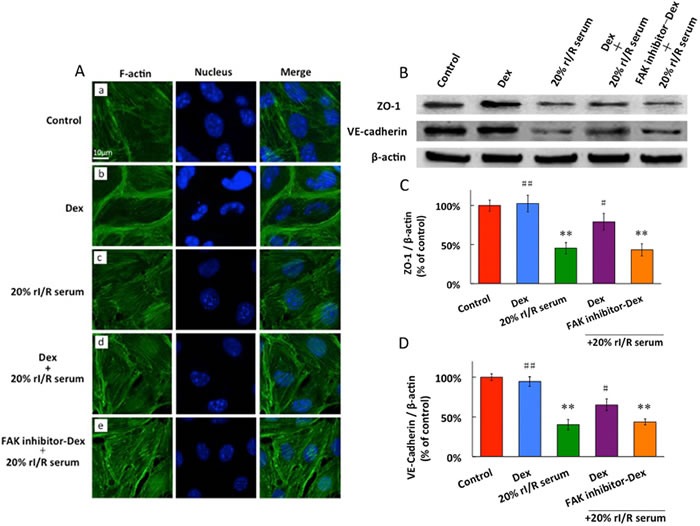
Figure 5. The effects of rI/Rserum on F-actin cytoskeletal assembly.

A., and the ZO-1 and VE-cadherin expressions in PMVECs (B, C, D). PMVECs incubated with 10μM FAK inhibitor for 3 hours before 0.1μM dexmedetomidine treatment for 20 minutes, followed by 20% rI/R serum stimulation for 60 minutes and F-actin was detected by Immunofluorescence. FITC-phalloidin fluorescence of F-actin cytoskeletal assembly with DAPI staining for the nucleus in PMVECs: A. No visible increase in stress fiber formation in the normal cell; B. dexmedetomidine significantly enhanced cortical cytoskeleton staining; C. 20% rI/R serum induced dissolution of cortical cytoskeleton and formation of prominent stress fiber and intercellular gaps; D. Pretreatment with dexmedetomidine prevented rI/R serum induced actin stress fiber formation, and diminished the paracellular gaps ; E. FAK inhibitor reversed the effect of dexmedetomidine. B. ZO-1 and C. VE-cadherin were detected by western blot. Data are expressed as the percentage of control (mean ± SD, n = 4);**p < 0.01vs.Control;#p < 0.05, ##p < 0.01vs.20% rI/R serum.
