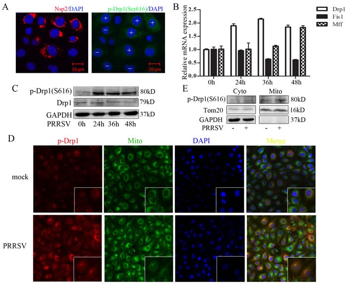
Figure 2. PRRSV infection enhances Drp1 phosphorylation and its mitochondrial translocation.

A. Immunofluorescence analysis showing the induction of drp1 Ser616 phosphorylation in PRRSV-infected cells. +, PRRSV-infected cells; -, uninfected cells. B.-C. Marc145 cells were infected with PRRSV for 0h, 24h, 36h, 48h, RNA and protein were collected for q-PCR and Weatern Blot analysis. D. Marc145 cells were infected with PRRSV for 36h and mitochondria were extracted. Phosphorylated Drp1 of subcellular fraction was determined by Western Blot. Cyto, cytosolic fraction; Mito, mitochondria fraction. Tom20, mitochondria marker; GAPDH, cytoplasmic marker. E. Marc145 cells were infected with PRRSV for 36h and location of phosphorylated Drp1was analyzed by confocal microscope. In the zoomed images, the yellow spots indicate the merge of phosphorylated Drp1 with mitochondria. Data are expressed as means with SEM of three independent experiments.
