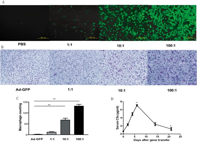
Figure 1. Expression of C5a protein in vitro and in vivo after adenoviral gene transfer.

A., Fluorescence images of HEK293 cells after transfection with phosphate buffered saline (PBS) or different multiplicities of infection (MOI) of adenovirus C5a (Ad-C5a) for 24 hr. B., Migration assay of chemotaxis of recombinant C5a to macrophages. HEK293 cells were transfected with Ad-GFP and different MOI (1:1, 10:1 and 100:1) of Ad-C5a. At 24 hr, the supernatants underwent trans-well assay. C., Quantitative analysis of trans-well assay. Data are mean ± SEM from 5 separate fields in each sample from 3 independent experiments. **P < 0.01. D., Detection of recombinant mouse C5a protein in ApoE−/− mouse plasma (n = 5). *P < 0.05 and **P < 0.01 vs day 0.
