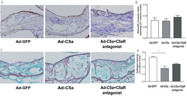
Figure 5. Collagen deposition and smooth muscle cell proliferation in mice.

A., Representative photomicrographs of atherosclerotic plaques stained for smooth muscle cells in the aortic root of ApoE−/− mice. B., Morphometric analysis of the stained areas for smooth muscle cells. C. Representative photomicrographs of atherosclerotic plaques stained for collagen in the aortic root of ApoE−/− mice.D., Morphometric analysis of the stained areas for collagen. Data are mean±SEM percentage positive area to total lesion area in 6 sections for each mouse. N = 8 per group. *P < 0.05.
