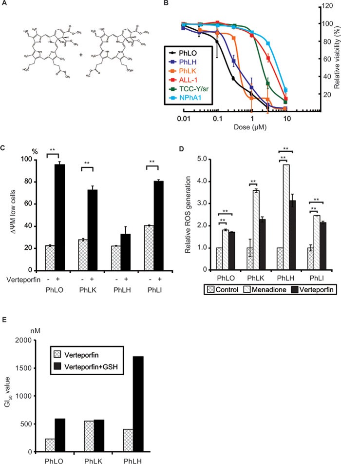
Figure 3. Verteporfin showed strong anti-leukemic effects through light-independent ROS production.

A. Chemical structure of verteporfin. B. Dose-dependent anti-leukemic effects of verteporfin on PDX cells and Ph+ ALL cell lines. PDX cells co-cultured with S17 cells and mono-cultured cell lines were treated with the indicated concentrations of verteporfin for 48 h. Viabilities were detected using a flow cytometer and presented as relative values to the viabilities of untreated cells. Each point represents the mean value for at least 3 independent experiments. Error bars show standard deviations. PDX cells were more sensitive to verteporfin than cell lines. C. Verteporfin reduced the mitochondrial membrane potential (ΔΨM) in PDX cells. The indicated PDX cells were treated with 1 μM verteporfin, labeled with JC-1 reagents, and analyzed with a flow cytometer. The ratio of low ΔΨM PDX cells was plotted on a bar graph. D. Verteporfin induced ROS production in PDX cells. The indicated PDX cells were incubated with verteporfin (2 μM) or menadione (50 μM) for 3 h. Menadione was used as the positive control of a ROS inducer. ROS production was measured by CellROX Green Oxidative Stress Reagents and plotted on a bar chart. E. GSH prevented the verteporfin-induced apoptosis in PDX cells. The indicated PDX cells co-cultured with S17 cells with or without 2 mM GSH in the culture medium were treated with the indicated dose of verteporfin as in B.. GI50 of verteporfin of the indicated PDX cells with or without GSH were determined and plotted on a bar chart. **: p < 0.001. GI50 were determined as results of at least 3 independent experiments. Error bars indicate standard deviations. GSH partly abolished the cytotoxicity of verteporfin in two of three PDX cells, suggesting that oxidative stress played a role in its cytotoxic effects.
