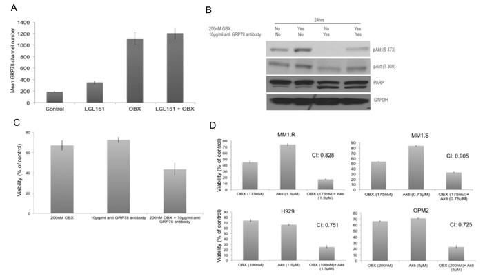
Figure 5.

A. MM1.R was left untreated or treated with 150nM OBX, 2μM LCL161 or the combination for 12hrs and cell surface expression of GRP78 was examined using flow cytometry. Y-axis represents mean GRP78 expression channel number. OBX has high auto fluorescence and hence results are presented as mean GRP78 expression channel number, which is the difference between mean channel number with GRP78 antibody and the mean channel number without GRP78 antibody for each treatment condition. Experiments were performed two times and error bars represent one standard deviation. B. MM1.R cells were treated with indicated concentrations of OBX, anti-GRP78 antibody (polyclonal antibody) or the combination and pAkt (S473), pAkt (T308) and PARP levels were examined by western blotting. GAPDH was used as a loading control. Experiments were performed two times and results from a representative experiment are shown. C. MM1.R cells were treated with indicated concentrations of OBX, anti GRP78 antibody (Clone C38) or the combination for 72hrs following which viability was measured by a MTT assay. OBX+GRP78 induced significantly more cell death than either of the agents alone (p < 0.05). The results presented are the mean of 5 independent experiments. D. MM1.R, MM1.S, H929 and OPM2 cells were treated with various concentrations of OBX for 72hrs, various concentrations of Akti for 48hrs or the drugs in combination. MTT assays were performed. Synergy was seen across multiple concentrations. The concentrations at which maximum synergy was observed is shown in the figure. Experiments were performed three times.
