
Figure 6. The anti-tumor effects of exogenously-pulsed B16-OVA vesicles on tumor growth.
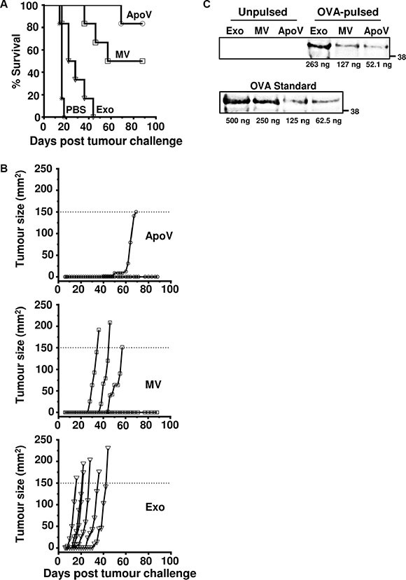
Mice were injected subcutaneously with ApoV, MV, Exo (isolated by differential centrifugation of ovalbumin-pulsed B16-OVA-derived vesicles), or PBS in the flank before challenged with B16-OVA cells at the opposite flank seven days later. Tumor size was recorded until tumors reached 150 mm2. (A) Percent survival of the four mouse groups (n = 6 mice/group). (B) Tumor size represented for individual mice. ApoV/Exo (P < 0.0005); ApoV/MV (not significant); MV/Exo (P < 0.0022); PBS/Exo (P < 0.004) by Mantel-Cox test. The protective results for ApoV were confirmed in an additional experiment. (C) OVA-pulsed Exo, MV or ApoV were probed by western blotting with rabbit anti-OVA and detected with donkey anti-rabbit IgG-DyLight-800. A titrated OVA standard was used to quantify the amount of OVA in each sample. Note, endogenously expressed ovalbumin in the B16-OVA cell line (‘Unpulsed’) is below the limit of detection by western blotting. One of two experiments performed.