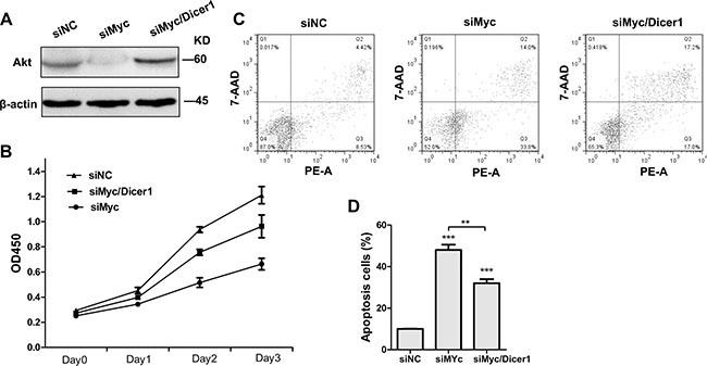
Figure 2. Depletion of Dicer1 abrogated the effect of c-Myc silencing in NB4 cells.

(A) Western blot of total AKT 48 h after the transfection with siRNAs agaist c-myc alone (siMyc) and together with siRNA against Dicer1 (siMyc/Dicer1). (B) Cell proliferation curve of NB4 cells transfected with the indicated siRNAs (n = 3, p < 0.05). (C) Apoptosis of NB4 cells treated with siNC, siMyc and siMyc/Dicer1 for 72 h as assessed by flow cytometry. PE-A on X-axis stands for PE-Annexin V. (D) Quantification of apoptosis cells shown in (C). Representative blot was shown from three independent experiments. Bar graph was represented as mean ± SD of three independent experiments performed in triplicates (n = 3). ** indicated P < 0.01 and *** indicated P < 0.001.
