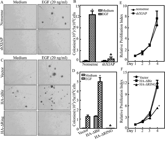
Figure 2. XIAP RING domain upregulated EGF induced bladder cell transformation.

A. and B. UROtsa(shXIAP) and UROtsa(Nonsense) cells were subjected to anchorage-independent assay in the presence or absence of EGF as indicated using the protocol described in the section of “Materials and Methods”. Representative images of colonies of UROtsa(shXIAP) and UROtsa(Nonsense) cells were captured under microscopy following 3 week incubation period; (A) the number of colonies was counted under microscopy and the results were presented as colonies per 50,000 cells from three independent experiments. The asterisk (*) indicates a significant increase in comparison to medium control and the symbol (♣) indicates a significant inhibition as compared with UROtsa(Nonsense) cells (p<0.05) (B). C. and D. UROtsa(Vector), UROtsa(HA-ΔBIR) or UROtsa(HA-ΔRING) cells were subjected to anchorage-independent assay in the presence or absence of EGF as indicated. Representative images of colonies of indicated cells were captured under microscopy following 3 week incubation period (C); the number of colonies was counted under microscopy and the results were presented as colonies per 50,000 cells from three independent experiments. The asterisk (*) indicates a significant increase as compared with UROtsa(Vector) cells, while the asterisk (♣) indicates a significant decrease as compared with UROtsa(Vector) cells (p<0.05) (D). E. and F. ATP assay to determine proliferation of cells was performed as indicated. The asterisk (*) indicates a significant inhibition as compared with UROtsa(Vector) cells.
