
Figure 4. XIAP RING domain inhibited p63α protein translation in human bladder epithelial cells.
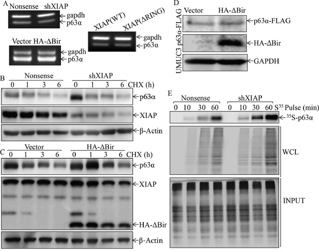
A. Total RNA was extracted from UROtsa transfectants or mouse primary bladder epithelial cells as indicated, then subjected to RT-PCR for evaluation of p63α mRNA expression. GAPDH was used as a loading control; B. and C. UROtsa(Nonsense) vs. UROtsa(shXIAP) cells (B), or UROtsa(Vector) vs. UROtsa(HA-ΔBIR) cells (C), were treated with 50 μg/ml cycloheximide (CHX) for the indicated times. The cell extracts were then subjected to Western Blot analyses of p63α protein degradation rates among the indicated cells. β-Actin was used as protein loading control. D. The cell extracts obtained from UMUC3(p63α-Flag/Vector) and UMUC3(p63α-Flag/HA-ΔBIR) cells were subjected to Western Blot to evaluate the effect of ectopic HA-ΔBIR expression on p63α-FLAG expression. E. Newly synthesized p63α protein in UROtsa(Nonsense) and UROtsa(shXIAP) cells was monitored by pulse assay using 35S-labeled methionine/cysteine as described in the section of “Materials and Methods”, WCL stands for whole cell lysate. Coomassie blue staining was used for protein loading control.