
Figure 5. XIAP RING domain initiated miR-4295 expression and in turn inhibited p63α mRNA 3′UTR activation.
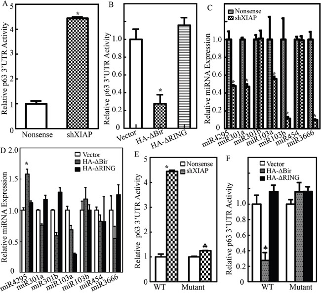
A. and B. Wild-type p63α 3′UTR luciferase reporters were co-transfected together with pRL-TK into UROtsa(Nonsense) vs. UROtsa(shXIAP) cells (A), or UROtsa(Vector) vs. UROtsa(HA-ΔBIR) and UROtsa(HA-ΔRING) cells (B), respectively. Twenty-four hours post transfection, the transfectants were extracted to evaluate the luciferase activity. TK was used as internal control. The results were presented as p63α 3′UTR activity relative to control vector transfectant, and each bar indicates a mean±SD from three independent experiments. The symbol (*) indicates a significant difference (P< 0.05); C. and D. The expression levels of miR-4295, miR-301a, miR-301b, miR-103a, miR-103b, miR-454 and miR-3666 were evaluated by real-time PCR as indicated. The results were normalized to U6; E. and F. UROtsa(Nonsense) vs. UROtsa(shXIAP) cells (E), or UROtsa(Vector) vs. UROtsa(HA-ΔBIR) and UROtsa(HA-ΔRING) cells (F), were transfected with either p63α 3′UTR luciferase reporter or mutant of p63α 3′UTR luciferase reporter that has an miR-4295 binding site mutation, Twenty-four hours post transfection, the transfectants were extracted to evaluate the luciferase activity. TK was used as internal control. The results were presented as p63α 3′UTR activity relative to control vector transfectant. Each bar indicates a mean±SD from three independent experiments. The symbol (*) shows a significant increase and the symbol (♣) indicates a significant inhibition (P< 0.05).