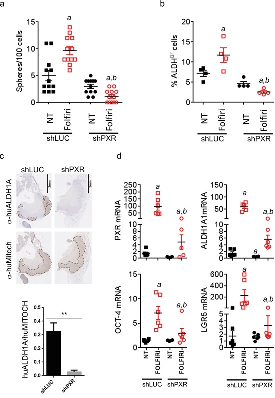
Figure 7. PXR depletion impairs chemotherapy-induced enrichment of PXR and CSC markers in vivo.

Tumor samples from mice sacrificed at day 48 (see Figure 6c) were processed for cell dissociation and, live (7-AAD-negative) tumor cells were further analyzed for their a. ability to initiate sphere formation in vitro and b. Aldefluor activity (% of ALDHbr cells). Data are expressed as mean±SEM. “a”, p<0.05 compared to non-treated shLUC tumors and “b”, p<0.05 compared to Folfiri-treated shLuc tumors. c. Hematoxylin/Eosin and immunostaining of Folfiri-treated shLuc and shPXR paraffin-embedded tumor sections (day 48) using antibodies directed against human ALDH1A (α-huALDH1A, detecting ALDH1A1 protein [54–56]) or human mitochondria (α-huMitoch). Ratios of ALDH1A/human mitochondria-stained areas on tumor sections (n=4/group) are presented below. Data are expressed as mean±SEM. **, p<0.005 d. Expression of the indicated mRNAs was quantified by RT-qPCR on cells isolated from dissociated tumors (day 48) of non-treated or Folfiri-treated mice. Data are expressed as mean±SEM. “a”, p<0.05 compared to non-treated shLUC tumors and “b”, p<0.05 compared to Folfiri-treated shLuc tumors.
