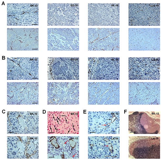
Figure 1. Histological appearance of BK-12, ED-15, HL-16, and LA-19 tumors.

A. Sections from donor patients' tumors stained with LYVE-1 (upper panels) or CD31 (lower panels). Bars, 100 μm (upper panels) or 200 μm (lower panels). B. Sections from patient-derived xenograft (PDX) tumors stained with LYVE-1 (upper panels) or CD31 (lower panels). Bars, 100 μm (upper panels) or 200 μm (lower panels). C. Adjacent sections from a BK-12 PDX tumor stained with LYVE-1 (upper panel) or CD31 (lower panel) showing that LYVE-1 and CD31 staining discriminated well between lymphatics and blood vessels. Bar, 25 μm. D. Images of a BK-12 PDX tumor showing functional intratumoral lymphatics. Upper panel, section stained with Prussian Blue showing many positively-stained low-diameter lymphatics (black arrows). Bar, 60 μm. Lower panel, section stained with LYVE-1 showing two positively-stained large-diameter ferritin-filled lymphatics (black arrows) and two unstained erythrocyte-containing blood vessels (red arrows). Bar, 30 μm. E. Sections from a BK-12 and an LA-19 PDX tumor stained with LYVE-1 showing intratumoral lymphatics with (red arrows) and without (black arrows) intraluminal tumor cells. Bar, 25 μm. F. Images showing metastatic growth in lymph nodes of mice bearing a BK-12 or an LA-19 PDX tumor. Bar, 650 μm.
