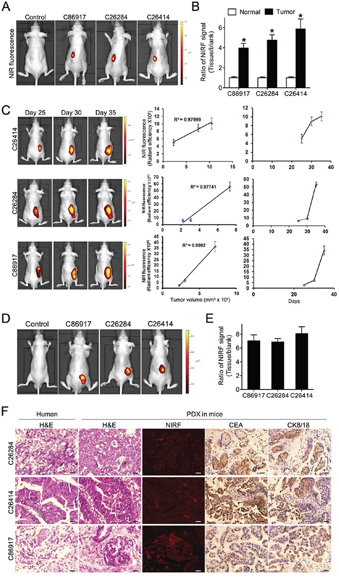
Figure 3. Uptake of NIRF dye and dye-drug conjugate in gastric cancer PDX models.

A. NIRF imaging of PDX models established by implanting 3 different human fresh gastric cancer specimens to one-side subrenal capsules of nude mice. Mice without implantation were used as control. Representative images are shown. B. Quantification of MHI-148 dye uptake in both normal kidney and subrenal tumor xenografts from PDX models. Data are presented as the ratio of dye uptake intensity as normalized to blank region (mean ± SD, n=5). *p<0.05. C. The correlation of tumor-emitting NIRF signal intensity with tumor size from 3 PDX models as measured at 3 different time points (mean ± SD, n=5). Representative images are shown. The mean was used to plot the correlation curves. D. NIRF imaging of IR-783-gemcitabine (NIRG) uptake in 3 different human gastric cancer PDX models. Mice without implantation were used as control. Representative images are shown. E. Quantification of NIRG dye-drug uptake in D. Data are presented as the ratio of dye uptake intensity as normalized to blank region (mean ± SD, n=5). F. H&E, NIRF and IHC analyses of tumor tissues derived from both PDX mouse models and original patient samples. Original magnification, ×400; scale bars represent 20 μm.
