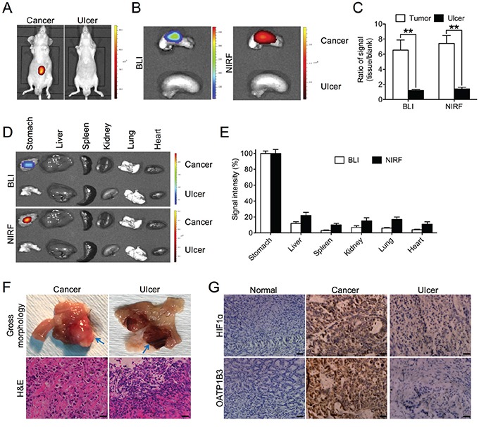
Figure 5. Preferential uptake of NIRF dye in gastric cancer relative to gastritic tissues.

A. In vivo NIRF imaging of mice bearing either orthotopic luc-tagged gastric tumor xenografts (left) or gastric ulcer (right). B. Ex vivo dual BLI/NIRF imaging of mouse stomach in A. C. Quantification of B for both modalities. Data are presented as the ratio (mean ± SD, n=5) of BLI/NIRF signal intensity as compared to blank region. **p<0.01. D. Ex vivo dual BLI/NIRF imaging of select organs, including the stomach, liver, spleen, kidney, lung and heart, as dissected from mice in A. E. Quantification of both BLI and NIRF signal intensity from the tumor-bearing experimental group in D. Data are presented as the percentage (mean ± SD, n=5) of signal intensity as normalized to stomach. Signal intensity in stomach is set as 100%. F. Gross morphology and H&E staining of gastric cancer and ulcer tissues in A. Representative images are shown. Blue arrows indicate tumor (left panels) and ulcer (right panels) area respectively. Original magnification, ×400; scale bars represent 20 μm. G. IHC analysis of HIF1α and OATP1B3 expression in gastric normal, cancer and ulcer tissues. Representative images are shown. Original magnification, ×400; scale bars represent 20 μm.
